Abstract
Vascular ring presenting as failure to thrive
Venkata Sushma Chamarthi MD, FAAP, DABOM, Sastry Chamarthi MD
Corresponding author: Venkata Sushma Chamarthi
Contact Information: Vchamarthi1@valleychildrens.org
DOI: 10.12746/swjm.v14i58.1603
ABSTRACT
A 6-month-old female presented with persistent post-prandial vomiting and poor weight gain despite adequate caloric intake. Physical examination revealed an emaciated but otherwise stable infant. Laboratory findings were normal. A barium esophagogram demonstrated posterior esophageal indentation suggestive of extrinsic compression, and computed tomography angiography revealed a right-sided aortic arch with retroesophageal Kommerell diverticulum and aberrant left subclavian artery – completing a vascular ring. Surgical division of the ligamentum arteriosum led to rapid symptom resolution and catch-up growth. This case underscores the importance of considering vascular rings in infants with unexplained feeding difficulties or failure to thrive, even in the absence of respiratory symptoms.
Keywords: Vascular ring, Kommerell diverticulum, Failure to thrive
CASE
A 6-month-old African American girl presented with persistent post-prandial vomiting and poor weight gain for two months. She had gained only 140 grams during this period (2 g/day) despite adequate caloric intake. Her parents described gagging and nasal regurgitation during feeding but denied respiratory distress, stridor, or recurrent infections. Physical examination revealed an emaciated but well-hydrated infant with weight below the 3rd percentile and otherwise normal cardiopulmonary findings.
Laboratory evaluation, including complete blood count, metabolic panel, thyroid function, and nutritional marker, unremarkable. A barium esophagogram demonstrated posterior indentation of the esophagus suggestive of extrinsic compression. Computed tomography angiography of the chest revealed a right-sided aortic arch with retroesophageal Kommerell diverticulum and aberrant left subclavian artery, completing a vascular ring via the ligamentum arteriosum (Figure 1). Echocardiography confirmed right-aortic-arch anatomy without intracardiac defects.
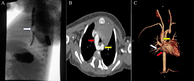
Figure 1. A: Lateral barium esophagogram showing a posterior indentation of the upper thoracic esophagus (white arrow) caused by external vascular compression. The smooth, crescent-shaped impression along the posterior wall is characteristic of a vascular ring. B: Axial contrast-enhanced CT chest image at the level of the aortic arch demonstrating a right-sided aortic arch with a retroesophageal Kommerell diverticulum (yellow arrow) giving rise to an aberrant left subclavian artery (red arrow) encircling the esophagus and trachea. C: Three-dimensional reconstructed CT angiogram confirming the complete vascular ring formed by the right aortic arch (white arrow), retroesophageal Kommerell diverticulum (yellow arrow), and ligamentum arteriosum (red arrow). The relationship of the vascular structures to the trachea (T) and esophagus (E) is clearly visualized, illustrating the site of compression relieved after surgical division.
The patient underwent surgical division of the ligamentum arteriosum via left thoracotomy. The postoperative course was uneventful. She showed rapid improvement in feeding tolerance with cessation of vomiting, and follow-up visits confirmed catch-up growth and resolution of feeding difficulties.
DISCUSSION
Vascular rings are rare congenital malformations of the aortic arch system that encircle and compress the trachea, esophagus, or both, accounting for <1 % of congenital cardiovascular anomalies.1 The most common symptomatic forms include double aortic arch and right aortic arch with aberrant left subclavian artery and Kommerell diverticulum.1,2 Most patients present with stridor, recurrent respiratory infections, or dysphagia; isolated gastrointestinal manifestations such as feeding difficulties and poor weight gain, are uncommon and may delay diagnosis.2 Compression limited to the esophagus rather than the airway can explain the absence of respiratory symptoms.
The diagnosis requires a high index of suspicion, particularly in infants with persistent vomiting or failure to thrive that does not respond to nutritional interventions. CT angiography and 3D reconstruction provide definitive visualization of the aortic arch and its relationship to the esophagus and trachea and allow precise surgical planning.1,3 Surgical division of the ligamentum arteriosum is curative in most cases, with rapid symptom resolution and excellent long-term outcomes.3
This case highlights the importance of being aware of vascular rings as a potential cause of unexplained feeding difficulties in infancy. Early recognition through targeted imaging allows timely intervention, prevents prolonged morbidity, and ensures optimal growth and development.
REFERENCES
- Backer CL, Mongé MC, Popescu AR, et al. Vascular rings. Semin Pediatr Surg. 2016;25(3):165–75.
- Depypere A, Proesmans M, Cools B, et al. The long-term outcome of an isolated vascular ring: a single-center experience. Pediatr Pulmonol. 2019;54(12):2028–34.
- Gikandi A, Chiu P, Crilley N, et al. Outcomes of patients undergoing surgery for complete vascular rings. J Am Coll Cardiol. 2024;84(14):1279–92. doi:10.1016/j.jacc.2024.05.078. PMID: 39322321.
Article citation: Chamarthi VS, Chamarthi S. Vascular ring presenting as failure to thrive. The Southwest Journal of Medicine. 2026;14(58):40–42
From: Valley Children’s Healthcare, Madera, CA (VSC) Clinica Sierra Vista Elm Community Health Cnter, Fresno, CA (SC)
Conflicts of interest: none
This work is licensed under a Creative Commons Attribution-ShareAlike 4.0 International License.