Abstract
Challenging diagnosis of pulmonary scedosporiosis in an immunocompetent elderly woman
Iqra Jabeen Mazhar MD, Taimoor Haider MD, Syed Asif Hussain MD, Ahmareen Khan MD
Corresponding author: Iqra Jabeen Mazhar
Contact Information: Iqrajabeen65@gmail.com
DOI: 10.12746/swjm.v14i61.1671
ABSTRACT
We report a rare case of pulmonary Scedosporium infection in a 64-year-old immunocompetent woman who presented with exertional dyspnea and intermittent wheezing. Imaging revealed a right lower lobe infiltrate, with a positron emission tomography (PET) scan showing hypermetabolic activity, raising concern for malignancy. CT-guided biopsy showed noncaseating granulomas that suggested a broad list of differential diagnosis. Eventually, bronchoalveolar lavage cultures identified Scedosporium species. The patient was managed with antifungal therapy. This case highlights the diagnostic challenges of fungal infections in immunocompetent individuals and the importance of considering opportunistic pathogens even in the absence of classical risk factors.
Keywords: Pulmonary scedosporium infection, immunocompetent host, noncaseating granulomatous lung disease
INTRODUCTION
The Scedosporium genus belongs to the filamentous fungi and comprises many species, with S. boydii and S. apiospermum being the most commonly isolated species.1 This genus is commonly found in the environment, particularly in soil, polluted water, and decaying vegetation, where it typically exists as a saprophyte. Scedosporium species are opportunistic pathogens that commonly affect individuals with immunocompromised conditions, such as hematological malignancies, solid organ transplant recipients, patients undergoing corticosteroid therapy, or prolonged neutropenia.2 However, they have also been reported in individuals without immunocompromised states, such as those with a history of near-drowning, major trauma, prior surgical history, or foreign body insertions.3,4
Scedosporium can cause a wide range of clinical manifestations, depending on the site of inoculation and host immunity, ranging from cutaneous infections, pulmonary infections, sinusitis, bone and joint infections, to disseminated infections.2
Pulmonary scedosporiosis is a particularly concerning manifestation that resembles other chronic infections or malignancies on imaging. Radiographic findings, such as infiltrates and nodules, may point toward other conditions like tuberculosis, chronic fungal infections, or lung malignancy. Due to the nonspecific presenting symptoms of chronic cough, dyspnoea, hemoptysis, and constitutional symptoms, diagnosis is often delayed and challenging. The culprit pathogen is identified with bronchoalveolar lavage (BAL) with fungal cultures, which is considered the gold standard but can be slow, or with molecular diagnostics.5 MALDI-TOF (Matrix-Assisted Laser Desorption/Ionization Time-of-Flight) is an alternative, rapid, and cost-effective method to isolate Scedosporium from the specimen.
We report a case of pulmonary scedosporiosis in an immunocompetent elderly woman who presented with nonspecific symptoms of dyspnoea on exertion and no systemic signs of infection. Further investigations revealed that Scedosporium can be diagnosed with BAL, although diagnosis can be challenging. This case places emphasis on the need to conduct a comprehensive workup when the clinical presentation is nonspecific, even in immunocompetent individuals.
CASE PRESENTATION
A 64-year-old woman with a history of Takotsubo/stress-induced cardiomyopathy after a motor vehicle collision and sleep apnea was seen in the pulmonology clinic for symptoms of dyspnea. The shortness of breath occurred on exertion, was more pronounced in the morning, and was associated with intermittent wheezing. She denied symptoms of fever, chills, weight loss, leg swelling, hemoptysis, orthopnea, and paroxysmal nocturnal dyspnea.
The patient is not immunocompromised but reports multiple episodes of pneumonia as a child and also had a remote history of smoking 2 packs per year. She lives in a home that is 100 years old but does not report any evidence of mold. Her pulmonary function tests (PFTs) were consistent with reversible small airway obstructive disease associated with increased airway resistance. She had a computed tomography (CT) scan of the thorax, which showed a macrolobulated mass-like density in the right lower lobe that was concerning for malignancy (Figures 1 and 2). A PET scan was obtained due to abnormal CT scan findings, and it redemonstrated a hypermetabolic right lower lobe mass most consistent with primary lung malignancy (Figures 3 and 4). This led to a CT-guided biopsy, which resulted in non-necrotizing granulomatous inflammation. Eventually, a decision was reached to conduct bronchoscopy with Bronchoalveolar Lavage (BAL) to form a diagnosis.
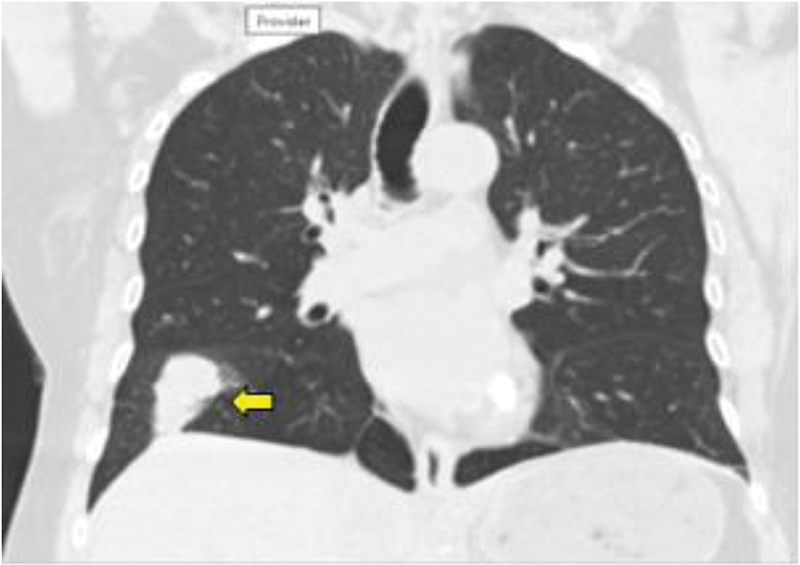
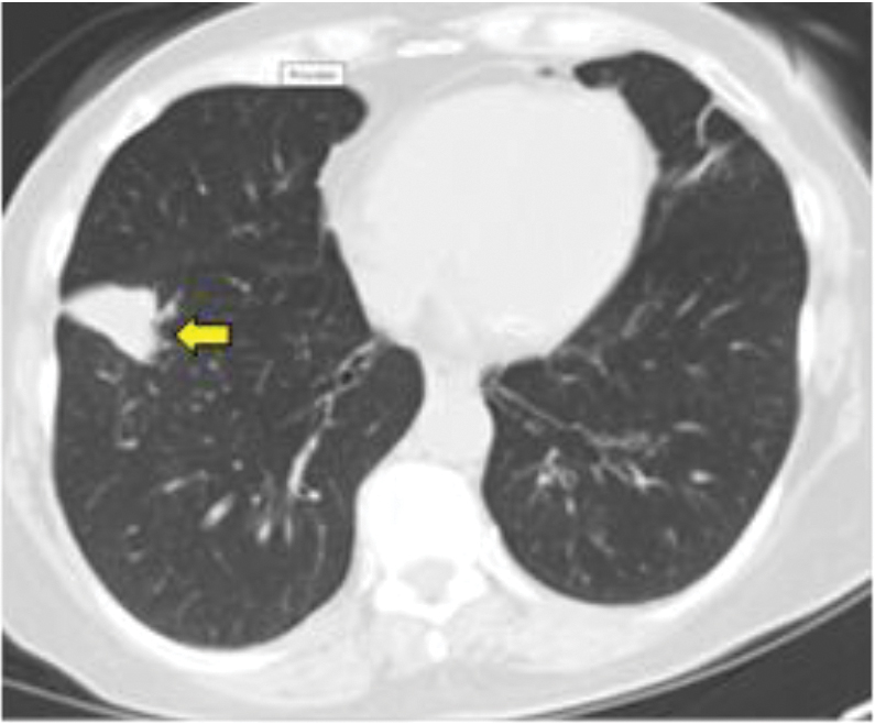
Figures 1 and 2. Computed Tomography (CT) scan of the thorax at presentation: Yellow arrow: Macrolobulated mass-like density in the right lower lobe. The persistence of this mass when compared to previous images raises concerns of malignancy.
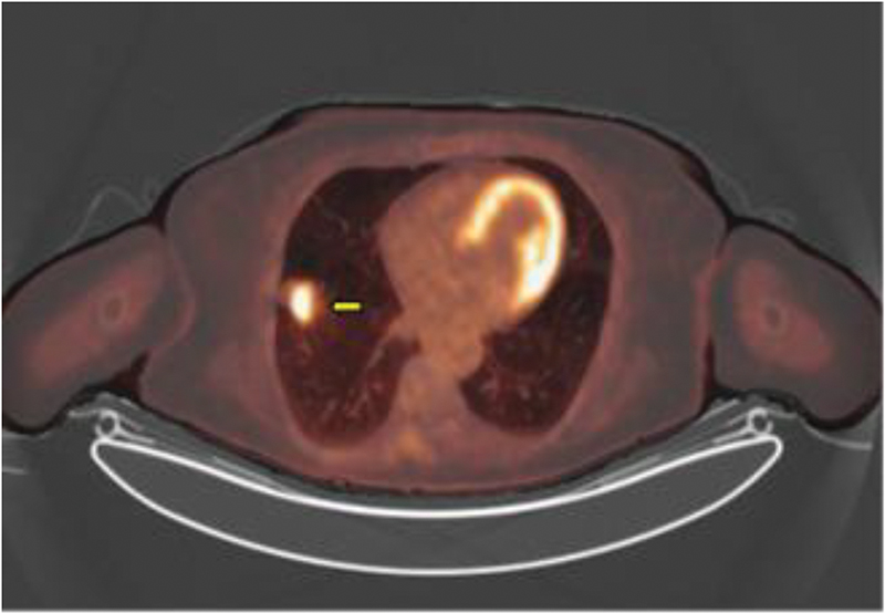
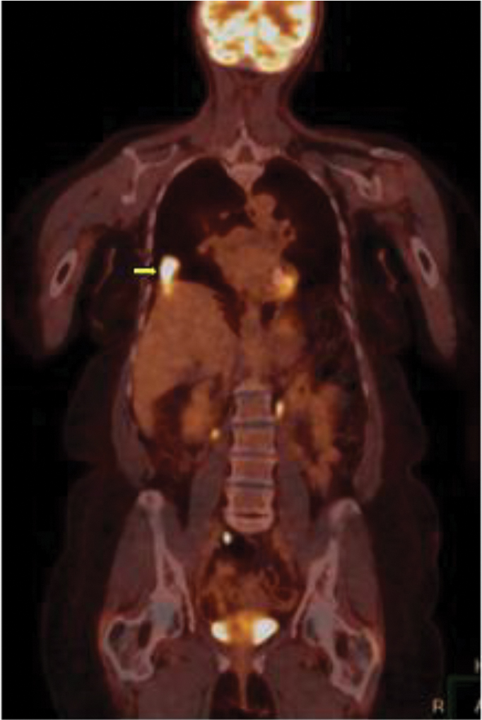
Figures 3 and 4. Positron Emission Tomography (PET) scan. Yellow arrow: Hypermetabolic right lobe mass that is most consistent with primary lung malignancy. No abnormal hilar or mediastinal uptake is identified.
The BAL fluid was sent for bacterial, fungal, and mycobacterial cultures, and these grew Scedosporium on fungal cultures. She was referred to the infectious disease department and was started on voriconazole 300 mg twice daily for Scedosporium pneumonia. Repeat CT scan almost 4 months after treatment showed a decrease in the right lower lobe mass-like density with residual disease (Figures 5, 6, and 7). She remains on voriconazole with a plan to repeat imaging and determine the duration of treatment based on her response.
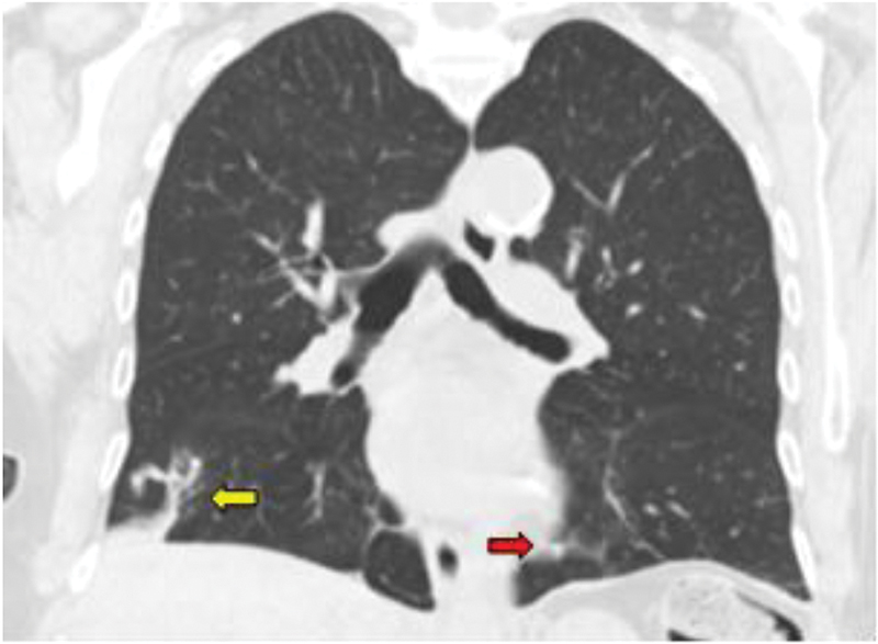
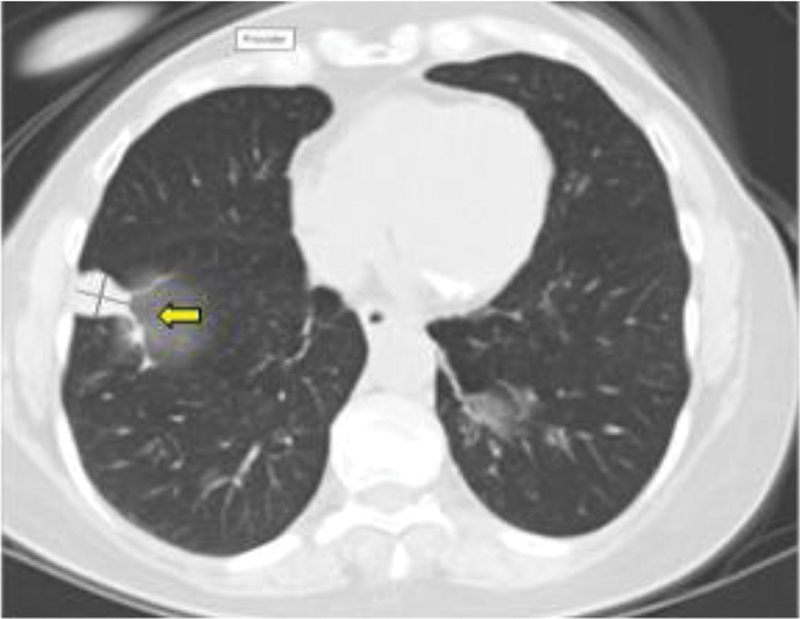
Figures 5 and 6. Computed Tomography scan of the thorax a year later, after treatment. Yellow arrow: A slight decrease is noticed in the right lobe mass-like density. Red arrow: Stable infiltrate and atelectasis of the inferior lingula.
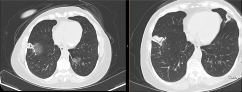
Figure 7. Computed Tomography scans of the thorax representing the difference in size of the right lobe mass after treatment with voriconazole.
CASE DISCUSSION
Scedosporium species are opportunistic pathogens that commonly affect people with immunodeficiency states, particularly individuals with hematological malignancies, solid organ transplantation, prolonged neutropenia, or those receiving corticosteroids.2 The infections can cause a spectrum of manifestations ranging from localized cutaneous infection to widespread, life-threatening disseminated disease. However, Scedosporium infection in an immunocompetent individual is very rare, which makes this case unique and diagnostically challenging.
This 64-year-old immunocompetent woman with a history of stress-induced cardiomyopathy presented with nonspecific respiratory symptoms that were not consistent with congestive heart failure. Her immunocompetence was confirmed by normal complete blood count (CBC) parameters and absence of neutropenia, negative HIV serology tests, and absence of immunosuppressive therapies. Our differential diagnosis included asthma and lung carcinoma.
Typical radiological findings in pulmonary Scedosporium present with patchy nodules or opacities involving one or more lobes.6 Lung cavities are another classic finding in chronic Scedosporium infections, which may be confused with tuberculosis or Aspergillus.7 The initial CT scan in this patient revealed a right lower lobe infiltrate; however, a subsequent PET scan showed hypermetabolic activity in the same area. This raised concerns for lung carcinoma; therefore, a CT-guided lung biopsy was scheduled. Histopathology revealed noncaseating granulomatous inflammation, which can be seen in various diseases, including tuberculosis, sarcoidosis, and fungal infections.8 However, granulomatous inflammation is a nonspecific feature, and differentiating among the possible pathologies remained a diagnostic challenge. Under the microscope, Scedosporium typically exhibits septate hyphae with distinctive branching patterns at irregular angles (>45 degrees).2 The conidiophores are often simple or branched, producing conidia that are oval or spherical and forming clusters at the end of hyphae.
Repeat imaging of this patient after five months showed persistent inflammation with signs of bronchiectasis and mucus plugging. This strengthened our suspicion of infectious etiology. In contrast to Aspergillus and Mucorales, which are rapidly progressive diseases and cause vascular invasion, Scedosporium tends to cause chronic, insidious infection with localized granulomatous lesions causing cavitation, bronchiectasis, and mucus plugging.5,6 Finally, a definitive diagnosis was reached after BAL, which produced a fungal culture growing Scedosporium species.
The diagnosis of Scedosporium was particularly difficult in this case due to nonspecific radiological and histopathological findings. However, BAL followed by culture remains the gold standard for diagnosing the infection, but advanced methods like MALDI-TOF mass spectrometry and internal transcribed spacer (ITS) sequencing can be used to identify this organism with specificity and sensitivity.5,9,10 MALDI-TOF identifies pathogens by analyzing their protein fingerprints through mass spectrometry, and ITS sequencing amplifies and sequences the fungal DNA’s internal transcribed spacer region. These newer and faster diagnostic modalities can provide rapid and precise identification of fungi at the species level.
Scedosporium possesses a high level of resistance to common antifungal drugs such as amphotericin B.2 However, itraconazole and voriconazole have shown positive treatment results for Scedosporium boydii infection, and focus has been placed on promptly treating S. boydii infection with these antifungals.1,3 Other treatment options include posaconazole and isavuconazole, but they have variable activity depending on the species. A residual lesion can be surgically resected after the initial response to voriconazole in order to reduce the risk of dissemination.7,11
CONCLUSION
The ubiquitous presence of Scedosporium in the environment and the immunocompromised state of an individual are typical causes of Scedosporium infection. However, it can also manifest in an immunocompetent person, and therefore, a broad differential should be considered when evaluating pulmonary infiltrates, particularly when the initial workup is inconclusive. The timely use of imaging and histopathology can lead to early recognition of uncommon fungal pathogens like this. Physicians should also be aware of the inherent resistance pattern of Scedosporium. This case also emphasizes the importance of prompt treatment with either voriconazole or itraconazole, which can improve patient outcomes. Increased awareness and reporting of such nonspecific presentations are essential to broaden our understanding of Scedosporium infections in immunocompetent hosts.
REFERENCES
- Lackner M, Klaassen CH, Meis JF, et al. Molecular identification tools for sibling species of Scedosporium and Pseudallescheria. Med Mycol. 2012;50(5):497–508. DOI: 10.3109/13693786.2011.618939
- Troke P, Aguirrebengoa K, Arteaga C, et al. Treatment of scedosporiosis with voriconazole: clinical experience with 107 patients. Antimicrob Agents Chemother. 2008;52(5):1743–50. DOI: 10.1128/AAC.01388-07
- Xiao Y, Li X, Hu L, et al. Clinical features and antifungal treatment of invasive Scedosporium boydii infection: report of a case and literature overview. Ann Clin Microbiol Antimicrob. 2024;23(1):94. DOI: 10.1186/s12941-024-00754-8
- Yao Y, Xu Q, Liang W, et al. Multi-organ involvement caused by Scedosporium apiospermum infection after near drowning: a case report and literature review. BMC Neurol. 2024;24(1):124. DOI: 10.1186/s12883-024-03637-9
- Cortez KJ, Roilides E, Quiroz-Telles F, et al. Infections caused by Scedosporium spp. Clin Microbiol Rev. 2008;21(1):157–97. DOI: 10.1128/CMR.00039-07
- Pellón Dabén R, Marco de Lucas E, Martín Cuesta L, et al. Imaging findings of pulmonary infection caused by Scedosporium prolificans in a deep immunocompromised patient. Emerg Radiol. 2008;15(1):47–9. DOI: 10.1007/s10140-007-0634-9
- Han J, Liang L, Li Q, et al. Diagnosis of pulmonary Scedosporium apiospermum infection from bronchoalveolar lavage fluid by metagenomic next-generation sequencing in an immunocompetent female patient with normal lung structure: a case report and literature review. BMC Infect Dis. 2024;24(1):308. DOI: 10.1186/s12879-024-09140-3
- Ohshimo S, Guzman J, Costabel U, et al. Differential diagnosis of granulomatous lung disease: clues and pitfalls: Number 4 in the Series “Pathology for the clinician” Edited by Peter Dorfmüller and Alberto Cavazza. Eur Respir Rev. 2017;26(145):170012. DOI: 10.1183/16000617.0012-2017
- Lackner M, de Hoog GS, Verweij PE, et al. Species-specific antifungal susceptibility patterns of Scedosporium and Pseudallescheria species. Antimicrob Agents Chemother. 2012;56(5):2635–42. DOI: 10.1128/AAC.05910-11
- Bernhard M, Zautner AE, Steinmann J, et al. Towards proteomic species barcoding of fungi – An example using Scedosporium/Pseudallescheria complex isolates. Fungal Biol. 2016;120(2):162–5. DOI: 10.1016/j.funbio.2015.07.001
- Masukane S, Kitahara Y, Okumoto J, et al. The effective treatment of lung infection due to scedosporium prolificans with voriconazole and surgery. Intern Med. 2017;56(8): 973–7. DOI: 10.2169/internalmedicine.56.7447
Article citation: Mazhar IJ, Haider T, Hussain SA, Khan A. Challenging diagnosis of pulmonary scedosporiosis in an immunocompetent elderly woman. The Southwest Journal of Medicine. 2026;14(61):85–90
From: Department of Internal Medicine, Shaukat Khanum Memorial Cancer Hospital and Research Center, Lahore, Pakistan (IM); Hospital Medicine, SSM Health St. Joseph Hospital, St. Charles, Missouri (TH, SH); Pulmonary Medicine, SSM Health St. Joseph Hospital, St. Charles, Missouri (AK)
Conflicts of interest: none
This work is licensed under a Creative Commons Attribution-ShareAlike 4.0 International License.