Abstract
Heavy metal exposure and women’s endocrine–metabolic disorders: An integrative review of PCOS, thyroid dysfunction, metabolic syndrome, and type 2 diabetes
Taslima Zaman MBBS, MPH, Nayla Mehereen Ahsan MBBS, Elora Shyama Tanni MBBS, MPH, Nowrin Islam MBBS, ABM, Fozlay Noor Shuvo MBBS, Purnota Saha MBBS, Abdullah Al Masud MD, FACP, Afrina Hossain Rimu MD, MS
Corresponding author: Afrina Hossain Rimu
Contact Information: Afrina.Rimu@yahoo.com
DOI: 10.12746/swjm.v14i61.1688
ABSTRACT
Cadmium (Cd), lead (Pb), mercury (Hg), and arsenic (As) are persistent environmental contaminants that can accumulate in human tissues and exert long-term biological effects. Although the acute toxicity of these metals is well established, increasing attention has focused on the effects of chronic, low-level exposure in the general population. These metals may disrupt endocrine and metabolic regulation through mechanisms including oxidative stress, mitochondrial dysfunction, immune activation, and interference with hormonal signaling pathways.
These mechanisms are relevant to endocrine and metabolic disorders affecting women, including polycystic ovary syndrome (PCOS), thyroid dysfunction, metabolic syndrome, and type 2 diabetes mellitus (T2DM). Hormonal transitions across the female life course may also influence responses to environmental exposures and disease expression.
This narrative review synthesizes epidemiological and mechanistic evidence published primarily between 2016 and 2024 and evaluates the relationships between exposure to Cd, Pb, Hg, and As and endocrine and metabolic disorders in women. Arsenic demonstrates the most consistent association with insulin resistance and T2DM. Cadmium has been associated with ovarian dysfunction, oxidative imbalance, and PCOS-related endocrine abnormalities. Lead and cadmium have also been linked to alterations in thyroid hormone levels, thyroid antibodies, and hypothyroid status. Findings regarding mercury remain less consistent.
Overall, current evidence supports a biologically plausible association between heavy metal exposure and the risk of endocrine and metabolic disorders in women. However, most available studies are observational. Longitudinal studies, standardized exposure assessments, and women-centered study designs are needed to clarify causal relationships and inform prevention strategies.
Keywords: Heavy metals, environmental exposure, polycystic ovary syndrome, thyroid dysfunction, metabolic syndrome, type 2 diabetes mellitus, women’s health
INTRODUCTION
Heavy metals are environmental contaminants found in air, water, soil, and food systems.1,2 Cadmium, lead, mercury, and arsenic are of particular concern because they persist in the environment, bioaccumulate, and may remain in human tissues for prolonged periods.3,4 These properties can result in continued internal exposure even when external exposure levels are low.
Earlier research focused primarily on acute poisoning and occupational exposure. More recent studies have examined the effects of chronic, low-level exposure in the general population. Even at relatively low levels, heavy metals may disrupt redox balance, mitochondrial function, endocrine signaling, and immune regulation.5,6 These effects may develop gradually and contribute to chronic endocrine and metabolic disorders.
The endocrine system is particularly sensitive to environmental disruption. Hormonal pathways depend on precise synthesis, transport, receptor binding, and feedback regulation. Heavy metals may interfere with several of these processes.7,8 Some effects may remain subclinical for years before contributing to clinically measurable disease.
Women may be uniquely vulnerable to the effects of heavy metal exposure because endocrine function changes across puberty, the reproductive years, pregnancy, lactation, and menopause. These transitions may alter exposure patterns, metal distribution, and biological responses.25,26 Nutritional status, iron balance, reproductive physiology, and metabolic differences may further influence susceptibility.
PCOS, thyroid dysfunction, metabolic syndrome, and T2DM are common endocrine and metabolic disorders affecting women. PCOS is associated with hyperandrogenism, ovulatory dysfunction, infertility, insulin resistance, and long-term cardiometabolic risk.10 Thyroid disorders are also more common in women and may affect reproductive, cardiovascular, and metabolic health. Metabolic syndrome and T2DM continue to increase globally and contribute substantially to the overall burden of disease.9
Genetic predisposition and lifestyle factors remain central to the development of these disorders. However, environmental exposures are increasingly recognized as potentially modifiable contributors. Understanding how heavy metals affect endocrine and metabolic pathways may help identify preventable risks and inform public health strategies.
This review synthesizes current evidence on the associations between heavy metal exposure and endocrine and metabolic disorders in women. It focuses on biological mechanisms, disease-specific associations, women-specific vulnerability, clinical implications, and research gaps.
METHOD
This narrative review synthesizes current evidence on the associations between heavy metal exposure and endocrine and metabolic disorders in women. Literature searches were conducted in PubMed, Scopus, and Google Scholar to identify relevant studies published between January 2016 and June 2024. Earlier landmark studies were also included when they provided important mechanistic insights or foundational evidence.
Search terms included combinations of “cadmium,” “lead,” “mercury,” “arsenic,” “heavy metals,” “environmental exposure,” “polycystic ovary syndrome,” “thyroid dysfunction,” “metabolic syndrome,” “insulin resistance,” and “type 2 diabetes mellitus.” Reference lists of relevant articles were also reviewed to identify additional studies.
The initial search retrieved more than 350 records. Titles and abstracts were screened for relevance, followed by full-text review of potentially relevant articles. Approximately 80 studies were included in the final synthesis.
Eligible studies included epidemiological, clinical, experimental, and review articles examining associations between heavy metal exposure and endocrine or metabolic outcomes. Greater emphasis was placed on studies with robust methodologies, larger sample sizes, prospective designs when available, and particular relevance to women’s health. Mechanistic studies were included to help elucidate potential biological pathways.
Because this was a narrative review, a formal risk-of-bias assessment was not performed. Instead, the evidence was interpreted by considering study design, consistency of findings, exposure assessment methods, and biological plausibility.
This review was intended to provide an overview of the current evidence and was not designed as a formal systematic review.
Table 1. Common Biomarkers of Heavy Metal Exposure and Associated Endocrine and Metabolic Effects in Women
This table summarizes commonly used biomarkers of heavy metal exposure and reported endocrine and metabolic effects. Reference values vary according to the biomarker, population, and agency guidance. For several metals, particularly lead, no clearly safe exposure level has been established.
| Metal |
Common biomarkers |
Common reference or guidance level |
Key endocrine–metabolic effects |
References |
| Arsenic (As) |
Urine, blood |
≤10 µg/L for drinking water |
Insulin resistance, impaired glucose metabolism, and type 2 diabetes mellitus (T2DM) |
11–14, 27 |
| Lead (Pb) |
Blood, bone |
No safe blood lead level established |
Thyroid dysfunction, hormonal imbalance |
19–22 |
| Mercury (Hg) |
Blood, hair |
Guideline-based; varies by mercury species and exposure source |
Dyslipidemia and metabolic alterations; evidence remains inconsistent |
23, 24 |
| Cadmium (Cd) |
Urine, blood |
Varies by biomarker and agency guidance |
PCOS-related features, oxidative stress, and insulin resistance |
15–18, 29 |
EXPOSURE PATHWAYS AND BIOACCUMULATION
Human exposure to heavy metals occurs through several pathways. Arsenic exposure is commonly associated with contaminated groundwater and, in some regions, contaminated food crops. Mercury exposure is primarily dietary, particularly through the consumption of fish and seafood containing methylmercury. Cadmium exposure is strongly associated with tobacco smoke and dietary sources. Lead exposure continues to occur through industrial contamination, aging infrastructure, older paint, and contaminated soil.1,2
After absorption, these metals differ in their distribution and retention within the body. Lead accumulates primarily in bone, where it can remain stored for decades. Cadmium accumulates mainly in the kidneys and liver and has a long biological half-life. Mercury distribution depends on its chemical form; methylmercury can cross biological membranes and accumulate in soft tissues, including the central nervous system. Arsenic is metabolized and excreted primarily in urine, although continuous exposure may sustain its biological effects.3,4
A major concern is that safe exposure thresholds remain uncertain for several metals, particularly lead. Even low-level exposure may contribute to cumulative biological effects over time. This is particularly relevant to endocrine and metabolic disorders, which often develop gradually through interactions among genetic, lifestyle, and environmental factors.
Biomonitoring methods vary according to the metal and exposure window being assessed. Blood levels often reflect recent exposure, whereas urinary measures may reflect cumulative body burden for some metals, such as cadmium. Bone lead measurements can provide an estimate of long-term exposure but are not commonly used in clinical practice. These differences are important when interpreting findings from epidemiological studies.
HEAVY METALS AND PCOS
PCOS is a complex endocrine disorder characterized by hyperandrogenism, ovulatory dysfunction, and metabolic abnormalities. Genetic predisposition and lifestyle factors remain central to its pathogenesis, although environmental exposures may also influence disease expression and severity.
Cadmium is among the environmental toxicants increasingly studied in relation to female reproductive health and PCOS-related phenotypes. Evidence from reproductive toxicology and epidemiological reviews suggests that cadmium may interfere with ovarian function, steroidogenesis, and follicular development.15,16 Cadmium-induced oxidative stress may also affect ovarian tissue and contribute to hormonal imbalance.6
Human studies suggest a possible association between cadmium exposure and PCOS-related endocrine features, although findings remain inconsistent. A prospective cohort study of healthy reproductive-aged women found that higher blood cadmium concentrations were associated with higher testosterone and anti-Müllerian hormone concentrations, as well as mild PCOS-like phenotypes.43 Another study of women with PCOS reported associations between heavy metal burden and insulin resistance, hirsutism, and other metabolic abnormalities.44 These findings do not establish causality but suggest that toxic metal exposure may modify disease severity in susceptible women.
Oxidative stress is a plausible mechanism linking cadmium exposure to reproductive dysfunction. Cadmium can increase reactive oxygen species, weaken antioxidant defenses, and impair mitochondrial function.6,45 Granulosa cells may be particularly vulnerable to oxidative injury, potentially affecting follicular maturation, oocyte quality, and steroid hormone production.46 Because oxidative stress and chronic inflammation are also implicated in PCOS, cadmium may exacerbate pre-existing endocrine and metabolic vulnerability rather than independently initiate PCOS.47
These findings have important reproductive and metabolic implications. Even subtle endocrine alterations may affect ovulation, fertility, and long-term cardiometabolic risk. Further longitudinal studies are needed to determine whether cadmium exposure contributes to PCOS progression or influences reproductive outcomes over time.44,45
HEAVY METALS AND METABOLIC SYNDROME
Metabolic syndrome is characterized by a cluster of metabolic abnormalities, including insulin resistance, central obesity, dyslipidemia, and hypertension. These conditions often occur together and increase the risk of T2DM and cardiovascular disease. Environmental exposures may also interact with dietary factors, obesity, inflammation, and genetic susceptibility in the development of metabolic syndrome.
Arsenic exposure has been consistently associated with impaired glucose metabolism and insulin resistance across diverse populations.11–14 Cadmium has also been linked to metabolic abnormalities, including altered lipid metabolism and inflammatory changes.18 Mercury exposure has been associated with dyslipidemia and metabolic syndrome in some studies, although the overall evidence remains inconsistent.23, 24
Several mechanisms may explain these associations. Heavy metals can increase oxidative stress, impair mitochondrial function, and promote low-grade inflammation.5,6 These processes may disrupt insulin signaling, lipid metabolism, and vascular function. However, metabolic syndrome is a multifactorial condition, and heavy metal exposure should be viewed as one potential contributor rather than a single primary cause.
HEAVY METALS AND TYPE 2 DIABETES
Among the metals reviewed, arsenic shows the most consistent association with T2DM. Epidemiological studies and reviews have linked arsenic exposure to insulin resistance, impaired glucose tolerance, and an increased risk of diabetes.11–14 Proposed mechanisms include oxidative stress, pancreatic β-cell dysfunction, altered insulin signaling, and inflammation. Cadmium may also contribute to glucose dysregulation, although the evidence is less consistent than that for arsenic. Some studies suggest that cadmium may affect β-cell function and insulin secretion, but findings vary according to population characteristics, exposure levels, and biomarker type.18
Mercury-related findings are more variable, and current evidence does not support a clear or consistent association between mercury exposure and T2DM.23,24
Overall, the available evidence indicates the strongest concern for arsenic in relation to T2DM. The association with cadmium remains biologically plausible but less certain, whereas mercury requires further investigation with improved exposure characterization.
HEAVY METALS AND THYROID DISORDERS
The thyroid gland is sensitive to environmental toxicants because thyroid function depends on tightly regulated hormone synthesis, iodine handling, and feedback mechanisms. Lead and cadmium exposure have been associated with alterations in thyroid hormone levels, thyroid antibodies, and hypothyroid status in population-based studies.19–22
These effects may occur through several mechanisms. Heavy metals may interfere with thyroid hormone synthesis, iodine utilization, thyroid peroxidase activity, and immune regulation.19–22,48,49 Cadmium may also contribute to oxidative stress and immune dysregulation relevant to thyroid autoimmunity.48 Although these mechanisms are biologically plausible, the available human evidence remains predominantly observational.
Women are disproportionately affected by thyroid disorders, especially autoimmune thyroid disease. This makes environmental thyroid disruptors an important consideration in women’s health. Hormonal fluctuations, immune differences, pregnancy, and menopause may further modify thyroid vulnerability. Future studies should examine whether the effects of heavy metal exposure on thyroid outcomes differ according to reproductive stage, iodine status, and autoimmune predisposition.
SHARED BIOLOGICAL MECHANISMS ACROSS HEAVY METAL EXPOSURE
Several biological mechanisms may link heavy metal exposure to endocrine and metabolic dysfunction. Oxidative stress is a central mechanism that disrupts redox balance and can damage proteins, lipids, and DNA.3,5,6 Mitochondrial dysfunction is closely related to oxidative stress and may impair energy metabolism and increase cellular vulnerability.6 Heavy metals may interfere with hormone synthesis, receptor signaling, and intracellular pathways.7,8 Cadmium and arsenic may be relevant to endocrine and metabolic disruption through these mechanisms.15–18 Inflammation may further contribute to metabolic dysfunction by impairing insulin signaling and promoting chronic metabolic stress.18 Immune dysregulation may also contribute to thyroid autoimmunity in association with lead and cadmium exposure.19–22
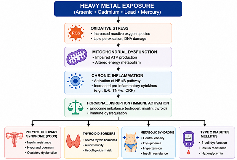
Figure 1. Proposed biological pathways linking heavy metal exposure to endocrine and metabolic disorders in women.
Heavy metals may contribute to endocrine and metabolic dysfunction through several interconnected mechanisms, including oxidative stress, mitochondrial dysfunction, inflammation, immune dysregulation, and disruption of hormonal signaling. These pathways may influence the development or progression of conditions such as PCOS, thyroid dysfunction, metabolic syndrome, and type 2 diabetes mellitus.
Table 2. Heavy Metals and Endocrine and Metabolic Disorders in Women: Proposed Mechanisms and Strength of Evidence
This table summarizes the major disorders discussed in this review, the metals most frequently implicated, proposed mechanisms, and the qualitative strength of the evidence. Strength of evidence was assessed narratively based on the consistency of epidemiological findings, biological plausibility, and the availability of mechanistic support.
| Disorder |
Metal(s) implicated |
Proposed mechanisms |
Strength of evidence |
References |
| PCOS |
Cadmium |
Oxidative stress, altered ovarian function, steroidogenic disruption, granulosa cell injury |
Moderate; mainly observational |
15–18, 43–47 |
| Thyroid dysfunction |
Lead, cadmium |
Altered thyroid hormone regulation, iodine pathway disruption, immune activation, thyroid antibodies |
Moderate |
19–22, 48, 49 |
| Metabolic syndrome |
Arsenic, cadmium, mercury |
Insulin resistance, mitochondrial dysfunction, inflammation, lipid disruption |
Moderate |
11–14, 18, 23, 24 |
| T2DM |
Arsenic, cadmium |
Impaired glucose metabolism, β-cell dysfunction, oxidative stress, altered insulin signaling |
Moderate-to-strong for arsenic; limited for cadmium |
11–14, 18 |
These pathways overlap and may help explain why different metals can affect multiple systems, including ovarian function, thyroid regulation, insulin sensitivity, and lipid metabolism. However, the same mechanisms may not apply equally across all metals or all women. Exposure dose and timing, nutritional status, and genetic susceptibility likely influence the resulting clinical outcomes.
DISCUSSION
This review supports a biologically plausible association between heavy metal exposure and endocrine and metabolic disorders in women. However, the strength of the evidence varies across metals and disease outcomes. Most available studies are observational, making causal relationships difficult to establish.
Among the metals reviewed, arsenic demonstrates the most consistent association with metabolic outcomes, including insulin resistance and T2DM.11–14 Epidemiological studies across diverse populations have repeatedly linked arsenic exposure to impaired glucose metabolism. Although residual confounding cannot be excluded, the consistency of these findings, together with supporting mechanistic evidence, strengthens the argument that arsenic may contribute to metabolic dysfunction.
Cadmium has emerged as another metal of concern. Evidence suggests that cadmium exposure may affect both reproductive and metabolic health. Studies examining PCOS-related outcomes have reported associations with hormonal alterations, insulin resistance, oxidative stress, and other metabolic abnormalities.15–18,43–47 However, these findings should be interpreted cautiously, as current evidence does not support a direct causal role for cadmium in the development of PCOS. Rather, cadmium may exacerbate existing endocrine and metabolic vulnerabilities in susceptible individuals.
The thyroid findings are also noteworthy. Population-based studies have associated lead and cadmium exposure with alterations in thyroid hormone levels, thyroid antibody concentrations, and hypothyroid status.19–22 Experimental evidence further suggests that cadmium may disrupt thyroid homeostasis through oxidative stress, immune dysregulation, and interference with iodine-dependent pathways.48,49 Because thyroid disorders disproportionately affect women, the potential influence of environmental exposures on thyroid function warrants greater attention in future endocrine research.
Mercury remains the metal with the least consistent evidence among those examined in this review. Some studies have reported associations between mercury exposure and dyslipidemia or metabolic syndrome, whereas others have found limited or no significant associations.23,24 Differences in exposure assessment, dietary habits, geographic variation, and the specific forms of mercury studied may partly account for these conflicting findings.
An important consideration is the complexity of real-world exposure patterns. Individuals are rarely exposed to a single environmental contaminant; instead, they encounter multiple chemicals simultaneously through food, drinking water, consumer products, occupational settings, and the surrounding environment. These exposures may interact through additive or synergistic mechanisms that are difficult to capture using traditional single-exposure models.36–38 The exposome framework provides a broader approach by considering cumulative environmental influences across the life course.38–40,50,51
Women-specific factors may further modify susceptibility to heavy metal toxicity. Pregnancy, lactation, and menopause are accompanied by physiological changes that can influence mineral metabolism and toxicant distribution. Lead stored in bone may be mobilized during periods of increased calcium demand, potentially increasing internal exposure even in the absence of recent environmental exposure.31,32 Iron deficiency may also enhance cadmium absorption, increasing vulnerability among women of reproductive age.33 These observations highlight the importance of considering sex-specific biological factors when evaluating environmental health risks.
Several limitations should be acknowledged. First, most human studies included in this review were cross-sectional, limiting the ability to establish temporal relationships between exposure and disease development. Second, exposure assessment methods varied substantially across studies. Biomarkers measured in blood, urine, and hair reflect different exposure windows and may not accurately represent long-term tissue accumulation. Third, variability in diagnostic criteria for conditions such as PCOS and metabolic syndrome may contribute to inconsistencies in findings across studies.
Future research should prioritize prospective cohort studies with repeated exposure measurements and standardized endocrine and metabolic outcomes. Greater attention should also be given to exposure mixtures, women-centered study designs, and vulnerable stages across the life course. Integrating environmental epidemiology with advances in biomonitoring, exposomics, and multi-omics technologies may improve understanding of the pathways linking environmental toxicants to endocrine and metabolic disorders.39,40,50,51
WOMEN-SPECIFIC VULNERABILITY ACROSS THE LIFE COURSE
Women may experience unique patterns of exposure and susceptibility because physiological transitions can influence both toxicant distribution and biological responses. Pregnancy, lactation, and menopause are accompanied by alterations in bone turnover and mineral metabolism that may affect the release and redistribution of stored metals. Lead provides a well-recognized example. Bone serves as a long-term reservoir for lead, and increased bone resorption during pregnancy and lactation may release previously stored lead into the circulation.31,32 Similar processes may also occur during menopause. As a result, cumulative lifetime exposure may be more relevant to health outcomes than measurements of recent exposure alone.
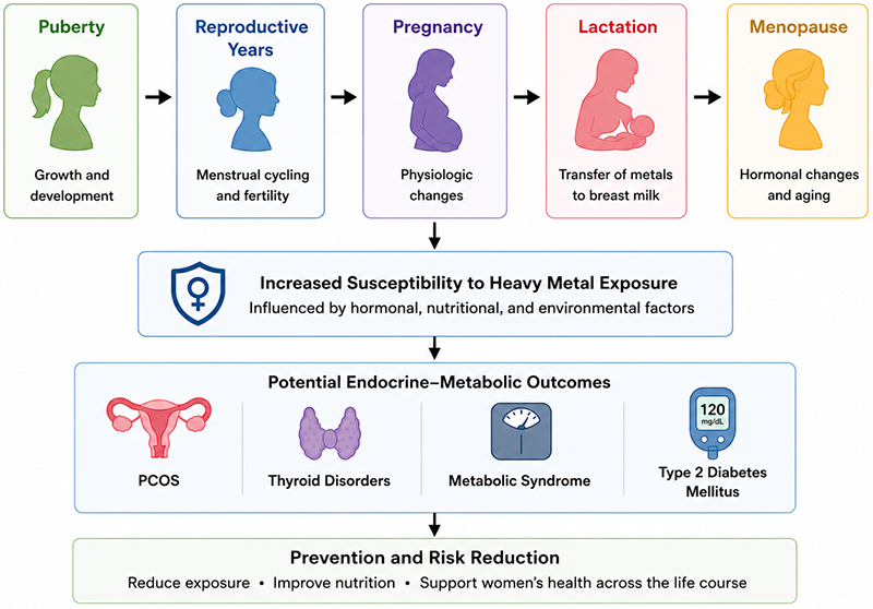
Figure 2. Women-specific susceptibility to heavy metal exposure across the life course.
Physiological changes during puberty, the reproductive years, pregnancy, lactation, and menopause may influence how women respond to heavy metal exposure. These factors may affect susceptibility to endocrine and metabolic disorders, including PCOS, thyroid dysfunction, metabolic syndrome, and type 2 diabetes mellitus. Reducing exposure and supporting women’s health throughout the life course may help reduce the risk of these disorders.
Micronutrient status may also influence toxic metal absorption. Iron deficiency has been associated with increased cadmium uptake because both(?) iron and cadmium share common intestinal transport mechanisms.33 Women of reproductive age are more susceptible to iron deficiency, which may increase vulnerability to cadmium accumulation. Deficiencies in calcium, zinc, and selenium may further affect toxicokinetics and toxicodynamic responses.34 Behavioral and social factors may also contribute to variability in exposure. Dietary practices, occupational factors, use of personal care products, and socioeconomic conditions can influence environmental exposure. Future studies should adopt a life-course perspective and incorporate reproductive history, nutritional status, and social determinants of health when evaluating exposure-related disease risk among women.35
MIXTURE TOXICOLOGY AND THE EXPOSOME PERSPECTIVE
Traditional environmental health research has often focused on individual chemicals. However, this approach may not adequately reflect real-world exposure scenarios. Most individuals encounter multiple contaminants simultaneously through air, water, food, consumer products, and occupational settings.
Combined exposures may produce additive, synergistic, or antagonistic biological effects.36 These interactions may influence oxidative stress pathways, mitochondrial function, endocrine signaling, and inflammatory responses.37 Consequently, evaluating individual chemicals in isolation may underestimate the overall environmental contribution to endocrine and metabolic disorders.
The exposome concept has emerged as a framework for understanding the cumulative impact of environmental exposures across the life course.38,39 This approach extends beyond chemical exposures to include behavioral, physical, and social determinants of health. Applying exposome-based methodologies to women’s endocrine and metabolic health may help identify susceptible populations and clarify exposure–disease relationships.
Recent advances in biomonitoring, data integration, and multi-omics technologies have expanded opportunities for exposome research.40,50,51 These approaches may enable more precise characterization of internal dose, biological responses, and individual susceptibility. Ultimately, exposome-informed studies may help inform prevention strategies and contribute to precision public health initiatives.
CLINICAL AND PUBLIC HEALTH IMPLICATIONS
Routine screening for heavy metal exposure is not currently recommended for most endocrine or metabolic disorders. However, environmental factors should not be overlooked during clinical assessment. In selected patients, obtaining a focused environmental exposure history may provide valuable information.
Clinicians may consider asking about occupational exposures, tobacco use, drinking water sources, dietary habits, residence near industrial areas, and exposure to older housing infrastructure.41 Identifying potential sources of exposure may help guide patient counseling and risk-reduction strategies.
From a public health perspective, prevention remains essential. Reducing environmental contamination, strengthening regulatory standards, and improving community awareness may help reduce the burden of exposure-related disease. Nutritional interventions aimed at correcting micronutrient deficiencies may represent an additional strategy for reducing susceptibility to toxic metal accumulation.34
Addressing environmental determinants of women’s health requires collaboration across multiple sectors. Healthcare professionals, researchers, policymakers, and public health agencies all have important roles in promoting safer environments and reducing preventable exposures. Continued investment in environmental surveillance and public health initiatives may help reduce disparities and improve long-term health outcomes among vulnerable populations.42
CONCLUSION
Heavy metal exposure represents a biologically plausible and potentially modifiable contributor to endocrine and metabolic disorders in women. Among the metals reviewed, arsenic shows the most consistent association with insulin resistance and T2DM.11–14 Cadmium has been linked to reproductive and metabolic disturbances, including endocrine features relevant to PCOS.15–18,43–47 Lead and cadmium have also been associated with alterations in thyroid hormone levels and thyroid autoimmunity.19–22 In contrast, evidence regarding mercury remains inconsistent.23,24
These findings should be interpreted with caution. Most available studies are observational and cannot establish causality. Differences in exposure assessment methods, study populations, and disease definitions further contribute to variability in findings across the literature. Nevertheless, the convergence of epidemiological observations and mechanistic evidence supports the biological plausibility of these associations.
From a clinical perspective, environmental exposures may represent an additional layer of risk beyond traditional genetic and lifestyle factors. Although routine heavy metal screening is not warranted for most endocrine and metabolic disorders, targeted environmental exposure history-taking may help identify potentially preventable exposures in selected patients.41 Greater awareness among healthcare professionals may facilitate earlier recognition of environmental contributors to disease risk.
The public health implications are equally important. Efforts aimed at reducing environmental contamination, strengthening regulatory oversight, promoting nutritional adequacy, and addressing environmental inequities may help reduce the burden of exposure-related disease.34,42 Because women experience unique physiological transitions throughout the life course, prevention strategies should account for sex-specific vulnerabilities and reproductive health considerations.
Several research gaps remain. Future investigations should prioritize prospective cohort studies with repeated exposure measurements and standardized endocrine and metabolic outcomes. Greater emphasis should also be placed on exposure mixtures, life-course approaches, and women-centered study designs. Integrating traditional epidemiology with emerging fields such as exposomics and multi-omics may provide deeper insight into the complex interactions between environmental toxicants and endocrine and metabolic health.39,40,50,51
In conclusion, heavy metals should be considered among the environmental factors that may influence endocrine and metabolic health in women. A clearer understanding of these relationships may help inform prevention efforts, improve risk assessment, and support more comprehensive approaches to women’s health across the lifespan.
REFERENCES
- Tchounwou PB, Yedjou CG, Patlolla AK, et al. Heavy metal toxicity and the environment. EXS. 2012;101:133–64.
- Jaishankar M, Tseten T, Anbalagan N, et al. Toxicity, mechanism and health effects of some heavy metals. Interdiscip Toxicol. 2014;7(2):60–72.
- Jomova K, Valko M. Advances in metal-induced oxidative stress and human disease. Toxicology. 2011;283(2–3):65–87.
- Agency for Toxic Substances and Disease Registry. Toxicological profile for arsenic, cadmium, lead, and mercury. Atlanta: U.S. Department of Health and Human Services; 2021.
- Valko M, Rhodes CJ, Moncol J, et al. Free radicals, metals and antioxidants in oxidative stress. Chem Biol Interact. 2006;160(1):1–40.
- Flora SJS, Mittal M, Mehta A. Heavy metal-induced oxidative stress and its reversal by chelation therapy. Indian J Med Res. 2008;128(4):501–23.
- Diamanti-Kandarakis E, Bourguignon JP, Giudice LC, et al. Endocrine-disrupting chemicals: an Endocrine Society scientific statement. Endocr Rev. 2009;30(4):293–342.
- Zoeller RT, Brown TR, Doan LL, et al. Endocrine-disrupting chemicals and public health protection. Endocr Rev. 2012;33(3):378–455.
- Grundy SM. Metabolic syndrome update. Circulation. 2016;134(5):e299–e313.
- Azziz R, Carmina E, Chen Z, et al. Polycystic ovary syndrome. Nat Rev Dis Primers. 2016;2:16057.
- Grau-Perez M, Kuo CC, Spratlen MJ, et al. The association of arsenic exposure with glucose metabolism and diabetes. Diabetes Care. 2018;41(3):e35–e36.
- Maull EA, Ahsan H, Edwards J, et al. Evaluation of the association between arsenic and diabetes: a National Toxicology Program workshop review. Environ Health Perspect. 2012;120(12):1658–70.
- Wang SL, Chang FH, Liou SH, et al. Inorganic arsenic exposure and its relation to metabolic syndrome. Environ Int. 2007;33(6):805–11.
- Chen Y, Ahsan H. Arsenic exposure and type 2 diabetes mellitus: a review. Curr Diab Rep. 2017;17(9):78.
- Kim JH, Park HY, Bae S, et al. Associations of blood cadmium levels with polycystic ovary syndrome in reproductive-aged women. Sci Total Environ. 2019;680:1–7.
- Pollack AZ, Ranasinghe S, Sjaarda LA, et al. Cadmium and reproductive health in women: a systematic review of the epidemiologic evidence. Curr Environ Health Rep. 2014;1(2):172–84.
- Heindel JJ, Blumberg B. Environmental obesogens and endocrine disruption. Endocrinology. 2019;160(1):10–19.
- Tinkov AA, Filippini T, Ajsuvakova OP, et al. Cadmium and metabolic syndrome: a review of epidemiological evidence. Environ Pollut. 2019;255:113232.
- Chen A, Kim SS, Chung E, et al. Thyroid hormones in relation to lead, mercury, and cadmium exposure in the National Health and Nutrition Examination Survey, 2007–2008. Environ Health Perspect. 2013;121(2):181–6.
- Dundar B, Oktem F, Arslan MK, et al. The effect of long-term low-dose lead exposure on thyroid function in adolescents. Environ Res. 2006;101(1):140–5.
- Nie X, Chen Y, Chen Y, et al. Lead and cadmium exposure, higher thyroid antibodies and thyroid dysfunction in Chinese women. Environ Pollut. 2017;230:320–8.
- Chen Y, Zhu C, Chen Y, et al. Are ethnic differences, urinary iodine status, lead and cadmium exposure associated with thyroid autoimmunity and hypothyroid status? BMJ Open. 2022;12(2):e050922.
- Moon SS. Association of mercury with metabolic syndrome. Diabetes Metab J. 2013;37(4):269–278.
- Park SB, Choi SW, Nam AY. Mercury exposure and metabolic outcomes: a systematic review. Environ Res. 2020;188:109734.
- Vahter M. Effects of arsenic on maternal and fetal health. Annu Rev Nutr. 2009;29:381–99.
- Kippler M, Tofail F, Gardner R, et al. Maternal cadmium exposure and child health outcomes. Environ Health Perspect. 2012;120(2):284–9.
- World Health Organization. Arsenic fact sheet. Geneva: World Health Organization; 2022.
- Centers for Disease Control and Prevention. National report on human exposure to environmental chemicals. Atlanta: Centers for Disease Control and Prevention; 2023.
- Agency for Toxic Substances and Disease Registry. Toxicological profile for cadmium. Atlanta: U.S. Department of Health and Human Services; 2021.
- Agency for Toxic Substances and Disease Registry. Toxicological profile for lead. Atlanta: U.S. Department of Health and Human Services; 2020.
- Gulson BL, Mizon KJ, Korsch MJ, et al. Mobilization of lead from human bone tissue during pregnancy and lactation—a summary of long-term research. Sci Total Environ. 2003;303(1–2):79–104.
- Silbergeld EK. Lead in bone: implications for toxicology during pregnancy and lactation. Environ Health Perspect. 1991;91:63–70.
- Åkesson A, Berglund M, Schütz A, et al. Cadmium exposure in pregnancy and lactation in relation to iron status. Am J Public Health. 2002;92(2):284–7.
- Zhai Q, Narbad A, Chen W. Dietary strategies for the treatment of cadmium and lead toxicity. Nutrients. 2015;7(1):552–71.
- Lewis RC, Meeker JD, Peterson KE, et al. Predictors of exposure to metals and associations with neurodevelopment among vulnerable populations: implications for women’s health research. Environ Res. 2018;164:507–15.
- Carlin DJ, Rider CV, Woychik R, et al. Unraveling the health effects of environmental mixtures: an NIEHS priority. Environ Health Perspect. 2013;121(1):A6–A8.
- Gore AC, Chappell VA, Fenton SE, et al. EDC-2: The Endocrine Society’s second scientific statement on endocrine-disrupting chemicals. Endocr Rev. 2015;36(6):E1–E150.
- Wild CP. Complementing the genome with an “exposome”: the outstanding challenge of environmental exposure measurement in molecular epidemiology. Cancer Epidemiol Biomarkers Prev. 2005;14(8):1847–50.
- Wild CP. The exposome: from concept to utility. Int J Epidemiol. 2012;41(1):24–32.
- Vermeulen R, Schymanski EL, Barabási AL, et al. The exposome and health: where chemistry meets biology. Science. 2020;367(6476):392–6.
- American College of Obstetricians and Gynecologists. Exposure to toxic environmental agents. Committee Opinion No. 832. Obstet Gynecol. 2021;138(1):e28–e37.
- World Health Organization. Chemicals, waste and pollution: preventing disease through healthy environments. Geneva: World Health Organization; 2023.
- Kim K, Pollack AZ, Nobles CJ, et al. Associations between blood cadmium and endocrine features related to PCOS phenotypes in healthy women of reproductive age: a prospective cohort study. Environ Health. 2021;20(1):64.
- Kirmizi DA, Baser E, Turksoy VA, et al. Are heavy metal exposure and trace element levels related to metabolic and endocrine problems in polycystic ovary syndrome? Biol Trace Elem Res. 2020;198(1):77–86.
- Massányi M, Massányi P, Stawarz R, et al. Evidence for ovarian and testicular toxicities of cadmium and their relevance to reproductive health. Reprod Med. 2021;2(1):1–20.
- Gao Y, Zou Y, Wu G, et al. Oxidative stress and mitochondrial dysfunction of granulosa cells in polycystic ovary syndrome. Front Med (Lausanne). 2023;10:1193749.
- Dabravolski SA, Nikiforov NG, Eid AH, et al. Mitochondrial dysfunction and chronic inflammation in polycystic ovary syndrome. Int J Mol Sci. 2021;22(8):3923.
- Buha A, Matović V, Antonijević B, et al. Overview of cadmium thyroid disrupting effects and mechanisms. Int J Mol Sci. 2018;19(5):1501.
- Ferrari SM, Fallahi P, Antonelli A, et al. Environmental issues in thyroid diseases. Front Endocrinol (Lausanne). 2017;8:50.
- Sdougkou K, Papazian S, Bonnefille B, et al. Longitudinal exposomics in a multiomic wellness cohort reveals distinctive and dynamic environmental chemical mixtures in blood. Environ Sci Technol. 2024;58(37):16302–16315.
- Maitre L, de Bont J, Casas M, et al. Multi-omics signatures of the human early life exposome. Nat Commun. 2022;13:7024.
Article citation: Zaman T, Ahsan NM, Tanni ES, Islam N, Shuvo FN, Saha P, Al Masud A, Rimu AH. Heavy Metal exposure and women’s endocrine–metabolic disorders: An integrative review of PCOS, thyroid dysfunction, metabolic syndrome, and type 2 diabetes. The Southwest Journal of Medicine. 2026;14(61):1–11
From: Southern Illinois University, Illinois, USA (TZ), Bangladesh Medical College, Dhaka, Bangladesh (NA), Sir Salimullah Medical College, Dhaka, Bangladesh (ET), Jahurul Islam Medical College, Kishoreganj, Bangladesh (NI), Satkhira Medical College, Satkhira, Bangladesh (FS), Dhaka Medical College, Dhaka,Bangladesh (PS), St. Louis University Hospital, St. Louis, Missouri,USA (AA), Department of Internal Medicine (Hospital Medicine), UF Health Leesburg Hospital, Florida, USA (AR).
Conflicts of interest: none
This work is licensed under a Creative Commons Attribution-ShareAlike 4.0 International License.