Abstract
Lipomatous hypertrophy of the interatrial septum: An unexpected finding complicating a routine CABG
Kiriti S. Vattikonda DO, MS, Courtney Barth BS, Christopher J. Peterson MD, MS, Michael S. Stump MD, John Lystash MD
Corresponding author: Christopher Peterson
Contact Information: Christopher.Peterson@unchealth.unc.edu
DOI: 10.12746/swjm.v14i61.1693
ABSTRACT
Lipomatous hypertrophy of the interatrial septum (LHIS) is a benign cardiac non-neoplastic mass. In the vast majority of cases, LHIS is an incidental finding found on cardiac imaging requiring no specific therapy. However, due to its size and location, LHIS can complicate cardiac surgery or structural heart interventional procedures.
Keywords: Lipomatous hypertrophy, interatrial septum, coronary artery bypass, cardiac mass, cardiac surgery
INTRODUCTION
Lipomatous hypertrophy of the interatrial septum (LHIS) is a benign cardiac non-neoplastic mass, often incidentally discovered on imaging and can often be followed without intervention. However, certain cases require surgical management. Here we present a challenging case of LHIS which complicated routine coronary artery bypass grafting.
CASE PRESENTATION
A 76-year-old man presented to a tertiary care center emergency department after experiencing two episodes of dull, non-radiating, central chest pain associated with diaphoresis over the past six hours. His past medical history included coronary artery disease (CAD) with remote stent placement 22 years prior in the left anterior descending (LAD) artery, recently diagnosed paroxysmal atrial fibrillation anticoagulated with apixaban, chronic obstructive pulmonary disease, diabetes mellitus type II, dyslipidemia, hypertension, obstructive sleep apnea, tobacco use disorder with smoking of 2 packs daily, and remote history of deep vein thrombosis 12 years prior. Vital signs on the initial presentation were stable without the patient requiring supplemental oxygen. On examination he was not in acute distress, cardiac auscultation revealed normal rate and regular rhythm without any clicks, rubs, gallops, or murmurs, and no obvious jugular venous distention was noted. Initial electrocardiogram was significant for sinus rhythm with pre-atrial contractions, poor R-wave progression, and non-specific ST elevation in V3. Initial high-sensitivity troponin and brain natriuretic peptide were 15 ng/ml and 29.5 pg/ml, respectively. A repeat troponin of 133 raised concern for acute coronary syndrome (ACS) and the patient was admitted to the cardiology service.
The initial working diagnosis for the patient’s presentation was non-ST elevation myocardial infarction which was supported by the results of transthoracic echocardiography (TTE) and coronary angiography described below. Transesophageal echocardiography (TEE) done before planned bypass showed an incidental mass in the right atrium with obstructive features. The differential diagnosis of the mass includes primary or secondary cardiac tumor, or endocarditis. The patient presentation, diagnostic testing, and pathologic testing were more suggestive of a primary cardiac tumor and less likely a secondary cardiac tumor or endocarditis given the absence of certain features seen in either.
Initial TTE demonstrated preserved ejection fraction of 60-65% with normal left ventricular systolic function and diastolic filling, mild aortic regurgitation, and a hypokinetic left apical septal segment. No obvious right atrial mass was visualized on TTE. The higher suspicion for ACS prompted left heart catheterization (LHC) revealing multivessel CAD with 70–80% diffuse stenosis of the proximal LAD, 95% in-stent restenosis of the mid-LAD, 90% stenosis of the ostial and proximal diagonal two branches, 80% stenosis of the mid left circumflex (LCX), 90% stenosis of ostial and proximal obtuse marginal branch 1 and obtuse marginal branch 2, 90% diffuse stenosis with two focal lesions of the distal LCX, 60% stenosis of the proximal right coronary artery, 70% stenosis of the proximal right coronary artery, 90% eccentric stenosis of the proximal right posterior descending artery. The LHC also showed low resting left ventricle end-diastolic pressure of 4 mmHg and pullback pressure gradient of 15–20 mmHg across the aortic valve. Cardiothoracic surgery was consulted for the multivessel CAD seen on LHC and planned for a double coronary artery bypass graft (CABG).
On periprocedural TEE, a previously unidentified large right atrial mass attached to the interatrial septum was noted, suspicious for severe LHIS with near obstruction of the superior vena cava (SVC) (Figures 1A and 1B, Videos 1 and 2). The cardiothoracic surgery team proceeded with CABG and resection of the right atrial mass was found to be necessary. Median sternotomy with left internal mammary harvest was performed. The Aorta was cannulated with a 24 French cannula; however, venous cannulation of the right atrial appendage was not possible given the presence of the obstructing mass. Alternatively, the SVC was dissected up to the innominate vein, and a 24 French angled cannula was placed. The inferior vena cava was cannulated with a 28 French-angled cannula to complete the cardiopulmonary bypass. Once the aorta was cross-clamped and cardioplegia was administered, coronary artery grafts were placed. The left internal mammary graft for the LAD and a radial artery graft, given a limited venous conduit, for the first OM branch of the LCX were used. The other LCX OM branches were noted to be small thus revascularization of the major territories was favored (Supplemental Figure 1). The right atrial free wall was then opened laterally to expose a large infiltrative mass emanating from the interatrial septum. Given the magnitude and infiltrative nature of the mass, an extensive partial resection was performed to achieve greater opening of the caval orifice (Figures 1C and 1D, Videos 3 and 4). Partial resection was favored as the primary focus of the operation was revascularization and palliative relief of the incidental obstruction. The patient’s age and underlying ischemic heart disease would have made the operation extremely complex and riskier if a total resection were pursued. An Aziyo intestinal submucosal patch measuring 8 cm × 5 cm was placed into the right atrial free wall defect in order to increase the circumference of the cavo-atrial junction and reduce SVC obstruction as autologous pericardium was not of adequate size. The patient was stabilized and transferred to the cardiac surgery intensive care unit. Grossly, the specimen consisted of multiple portions of adipose tissue ranging from 0.7 cm up to 2.3 cm in greatest dimension. Microscopic examination of the resected right atrial mass showed numerous histologic sections demonstrating portions of myocardium admixed with adipose tissue characteristic of LHIS (Figures 2A and 2B).
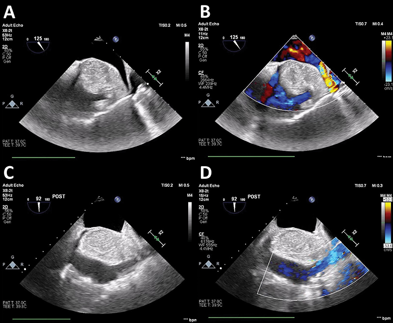
Figure 1. Pre-and Post-Operative Echocardiography of LHIS Mass TEE showing. (A) Pre-resection images of LHIS measuring 3.52 cm, sparing the fossa ovalis and obstructing the SVC, (B) Pre-resection doppler flow showing turbulent flow of SVC and right upper pulmonary vein, (C) partial post-resection of LHIS showing opening of the SVC, and (D) Partial post-resection doppler flow showing reduction of turbulent flow across SVC.
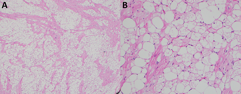
Figure 2. Histopathology Sections of LHIS Mass at 5x and 20x Hematoxylin and eosin stain of LHIS mass at (A) 5x magnification showing a patchwork of mature adipose tissue and hypertrophic myocytes and (B) higher power (20x) magnification demonstrating a mix of mature adipocytes and brown fat cells (vacuolated adipocytes).
DISCUSSION
Lipomatous hypertrophy of the interatrial septum is an uncommon hyperplasia of atrial septal fat first identified in 1964.1 Also known as lipomatous hypertrophy of atrial septum, its name is a misnomer as it actually involves hyperplasia of adipocyte cells,2 though this name has nevertheless persevered. Although LHIS represents abnormal hypertrophy, it is not a malignant growth but rather hyperplasia of mature adipose cells and brown fetal adipose cells.3 The incidence is between 1 and 8%1 and, though historically identified on autopsy, modern imaging techniques such as echocardiography, MR, and positron emission tomography-computed tomography (PET-CT) make diagnosis possible without a tissue sample.4 Its characteristic bilobar or dumbbell-shaped appearance ( sparing of the fossa ovalis), and septal thickness of >2 cm is considered diagnostic.3,4 Imaging of the mass should be reviewed by experts familiar with LHIS to avoid misdiagnosis and potential unnecessary surgical intervention.5 To this point, LHIS can have metabolically active brown fat with increased uptake on PET-CT imaging, which may be misinterpreted as an infectious or malignant lesion.6,7
Lipomatous hypertrophy of the interatrial septum is usually asymptomatic and often incidentally identified.8 However, its association with supraventricular arrhythmias and sudden cardiac death,8 possibly by disruption of cardiac myocytes,1 may also prompt identification. The association with advanced age and obesity suggests involvement of a metabolic condition or age-related epicardial fat accumulation, though this may also be confounding making it difficult to prove causation.8,9 Lipomatous hypertrophy of the interatrial septum typically does not require treatment, though surgical resection may be required for intractable symptoms, severe arrhythmias, right atrial obstruction, or compression of adjacent structures.9,10 Several differential pathologies should be considered, such as lipoma or myxoma, though these can often be differentiated due to the absence of a capsule, sparing of the fossa ovalis, fatty composition (confirmed on CT or MRI), absence of a stalk, and lack of rapid growth.3,8 In this case, there was no prior imaging that identified the mass, and it was discovered incidentally periprocedurally on TEE. The presence of the mass affected the surgical procedure, requiring cannulation at the innominate vein instead of the RA due to obstruction from the mass, and it was ultimately resected due to its size and degree of infiltration. After discharge, the patient was seen in the Cardiology and Cardiothoracic Surgery clinics reporting improved functional and activity tolerance. The patient had no further admissions or major adverse events after hospital discharge.
This case highlights that hypertrophy of the interatrial septum must be included in the differential diagnosis of any right atrial mass. Location, dumbbell shape, and sparing of the fossa ovalis are important clues in making the correct diagnosis. Surgical excision of LHIS is reserved for patients who have SVC obstruction as this case illustrates.
VIDEOS
Video 1: Pre-resection echocardiography film of lipomatous hypertrophy of the atrial septum
Video 2: Pre-resection Doppler echocardiography film flow showing turbulent flow of SVC and right upper pulmonary vein
Video 3: Partial post-resection echocardiography film of lipomatous hypertrophy showing the opening of the SVC
Video 4: Partial post-resection echocardiography film Doppler flow showing reduction of turbulent flow across SVC
REFERENCES
- Heyer CM, Kagel T, Lemburg SP, et al. Lipomatous hypertrophy of the interatrial septum: a prospective study of incidence, imaging findings, and clinical symptoms. Chest. 2003;124(6):2068–73. DOI: 10.1378/chest.124.6.2068
- Silbiger JJ, Bazaz R, Trost B. Lipomatous hypertrophy of the interatrial septum revisited. J Am Soc Echocardiogr. 2010;23(7):789–90. DOI: 10.1016/j.echo.2010.04.015
- Laura DM, Donnino R, Kim EE, et al. Lipomatous atrial septal hypertrophy: a review of its anatomy, pathophysiology, multimodality imaging, and relevance to percutaneous interventions. J Am Soc Echocardiogr. 2016;29(8):717–23. DOI: 10.1016/j.echo.2016.04.014
- O’Connor S, Recavarren R, Nichols LC, et al. Lipomatous hypertrophy of the interatrial septum: an overview. Arch Pathol Lab Med. 2006;130(3):397–9. DOI: 10.5858/2006-130-397-LHOTIS
- Nadra I, Dawson D, Schmitz SA, et al. Lipomatous hypertrophy of the interatrial septum: a commonly misdiagnosed mass often leading to unnecessary cardiac surgery. Heart. 2004;90(12):e66. DOI: 10.1136/hrt.2004.045930.
- Malik SB, Kwan D, Shah AB, et al. The right atrium: gateway to the heart—anatomic and pathologic imaging findings. Radio Graphics. 2015;35(1):14–31. DOI: 10.1148/rg.351130010
- Callaud A, Metrard G, Besse H, et al. Extinction of lipomatous hypertrophy of the interatrial septum FDG uptake with propranolol premedication. Clin Nucl Med. 2023;48(6):536–7. DOI: 10.1097/RLU.0000000000004640
- Burke AP, Litovsky S, Virmani R. Lipomatous hypertrophy of the atrial septum presenting as a right atrial mass. Am J Surg Pathol. 1996;20(6):678–85. DOI: 10.1097/00000478-199606000-00004
- Lampropoulos KM, Kotsas D, Iliopoulos T. Lipomatous hypertrophy of interatrial septum. BMJ Case Rep. 2012;2012:bcr2012006611. DOI: 10.1136/bcr-2012-006611
- Ianni U, Tofoni P, Molisana et al. Lipomatous atrial septal hypertrophy, an unexpected giant interatrial mass: a case report and literature review. J Cardiovasc Echogr. 2023;33(1):36–9. DOI: 10.4103/jcecho.jcecho_58_22
Article citation: Vattikonda KS, Barth C, Peterson CJ, Stump MS, Lystash J. Lipomatous hypertrophy of the interatrial septum: An unexpected finding complicating a routine CABG. The Southwest Journal of Medicine. 2026;14(61):110–114
From: Department of Internal Medicine, Virginia Tech Carilion School of Medicine, Roanoke, VA (KSV, CJP) Virginia Tech Carilion School of Medicine, Roanoke, VA (CB) Department of Pathology, Carilion Clinic, Roanoke, VA (MSS) Department of Cardiology, Virginia Tech Carilion School of Medicine, Roanoke, VA (JL)
Conflicts of interest: none
This work is licensed under a Creative Commons Attribution-ShareAlike 4.0 International License.